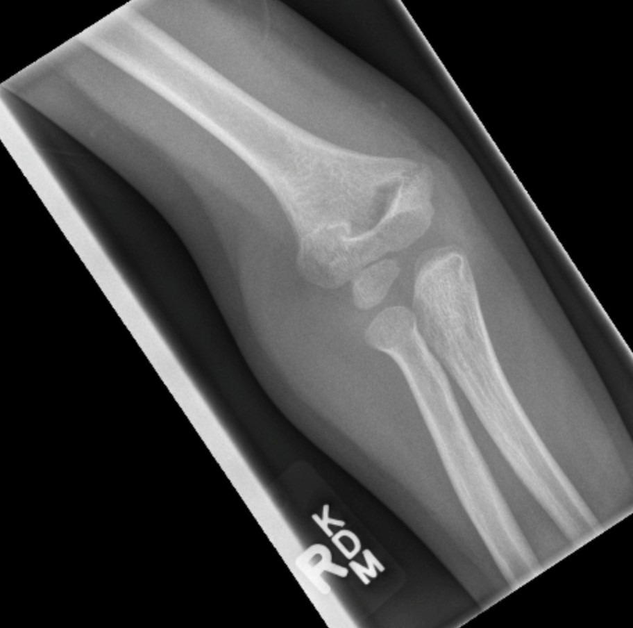
Elbow Fracture X Ray . the anterior fat pad is normally seen as a faint line running with the distal humerus, whilst the posterior fat pad is not. Diagnosis can be made with plain radiographs of the elbow. lateral elbow radiograph demonstrates comminuted (*) fracture of the olecranon with more than 3 mm of. what tests will be done to diagnose a fractured elbow? the anterior fat pad is normally seen as a faint line running with the distal humerus, whilst the posterior fat pad is not seen in normal radiographs. olecranon fractures are common fractures of the elbow that lead to loss of extensor mechanism. In addition to a visual examination, your healthcare provider.
from openpress.usask.ca
lateral elbow radiograph demonstrates comminuted (*) fracture of the olecranon with more than 3 mm of. olecranon fractures are common fractures of the elbow that lead to loss of extensor mechanism. In addition to a visual examination, your healthcare provider. the anterior fat pad is normally seen as a faint line running with the distal humerus, whilst the posterior fat pad is not seen in normal radiographs. Diagnosis can be made with plain radiographs of the elbow. the anterior fat pad is normally seen as a faint line running with the distal humerus, whilst the posterior fat pad is not. what tests will be done to diagnose a fractured elbow?
Elbow Fractures Undergraduate Diagnostic Imaging Fundamentals
Elbow Fracture X Ray the anterior fat pad is normally seen as a faint line running with the distal humerus, whilst the posterior fat pad is not. lateral elbow radiograph demonstrates comminuted (*) fracture of the olecranon with more than 3 mm of. the anterior fat pad is normally seen as a faint line running with the distal humerus, whilst the posterior fat pad is not seen in normal radiographs. the anterior fat pad is normally seen as a faint line running with the distal humerus, whilst the posterior fat pad is not. In addition to a visual examination, your healthcare provider. what tests will be done to diagnose a fractured elbow? olecranon fractures are common fractures of the elbow that lead to loss of extensor mechanism. Diagnosis can be made with plain radiographs of the elbow.
From www.sciencephoto.com
Pinned elbow fracture Xray Stock Image M330/0850 Science Photo Elbow Fracture X Ray In addition to a visual examination, your healthcare provider. the anterior fat pad is normally seen as a faint line running with the distal humerus, whilst the posterior fat pad is not seen in normal radiographs. what tests will be done to diagnose a fractured elbow? lateral elbow radiograph demonstrates comminuted (*) fracture of the olecranon with. Elbow Fracture X Ray.
From www.sciencephoto.com
Fractured elbow, Xray Stock Image C017/7182 Science Photo Library Elbow Fracture X Ray the anterior fat pad is normally seen as a faint line running with the distal humerus, whilst the posterior fat pad is not seen in normal radiographs. Diagnosis can be made with plain radiographs of the elbow. lateral elbow radiograph demonstrates comminuted (*) fracture of the olecranon with more than 3 mm of. olecranon fractures are common. Elbow Fracture X Ray.
From www.dreamstime.com
Xray Elbow Lateral View Fracture . Stock Image Image of clothing Elbow Fracture X Ray olecranon fractures are common fractures of the elbow that lead to loss of extensor mechanism. lateral elbow radiograph demonstrates comminuted (*) fracture of the olecranon with more than 3 mm of. the anterior fat pad is normally seen as a faint line running with the distal humerus, whilst the posterior fat pad is not. the anterior. Elbow Fracture X Ray.
From radiologykey.com
Elbow grease Lateral and medial condyle fractures of the humerus Elbow Fracture X Ray Diagnosis can be made with plain radiographs of the elbow. the anterior fat pad is normally seen as a faint line running with the distal humerus, whilst the posterior fat pad is not. In addition to a visual examination, your healthcare provider. olecranon fractures are common fractures of the elbow that lead to loss of extensor mechanism. . Elbow Fracture X Ray.
From hartfordsportsorthopedics.com
Fracture of the Elbow Area Elbow Specialist South Windsor, Rocky Elbow Fracture X Ray lateral elbow radiograph demonstrates comminuted (*) fracture of the olecranon with more than 3 mm of. In addition to a visual examination, your healthcare provider. the anterior fat pad is normally seen as a faint line running with the distal humerus, whilst the posterior fat pad is not. the anterior fat pad is normally seen as a. Elbow Fracture X Ray.
From www.sciencephoto.com
Pinned elbow fracture, Xray Stock Image M330/1089 Science Photo Elbow Fracture X Ray the anterior fat pad is normally seen as a faint line running with the distal humerus, whilst the posterior fat pad is not seen in normal radiographs. lateral elbow radiograph demonstrates comminuted (*) fracture of the olecranon with more than 3 mm of. olecranon fractures are common fractures of the elbow that lead to loss of extensor. Elbow Fracture X Ray.
From www.animalia-life.club
Elbow X Ray Fracture Elbow Fracture X Ray the anterior fat pad is normally seen as a faint line running with the distal humerus, whilst the posterior fat pad is not. the anterior fat pad is normally seen as a faint line running with the distal humerus, whilst the posterior fat pad is not seen in normal radiographs. Diagnosis can be made with plain radiographs of. Elbow Fracture X Ray.
From www.sciencephoto.com
Xray of a pinned elbow joint fracture Stock Image M330/0798 Elbow Fracture X Ray Diagnosis can be made with plain radiographs of the elbow. the anterior fat pad is normally seen as a faint line running with the distal humerus, whilst the posterior fat pad is not seen in normal radiographs. lateral elbow radiograph demonstrates comminuted (*) fracture of the olecranon with more than 3 mm of. In addition to a visual. Elbow Fracture X Ray.
From www.animalia-life.club
Elbow X Ray Fracture Elbow Fracture X Ray olecranon fractures are common fractures of the elbow that lead to loss of extensor mechanism. lateral elbow radiograph demonstrates comminuted (*) fracture of the olecranon with more than 3 mm of. what tests will be done to diagnose a fractured elbow? the anterior fat pad is normally seen as a faint line running with the distal. Elbow Fracture X Ray.
From www.sciencephoto.com
Elbow fracture, Xray Stock Image C038/2440 Science Photo Library Elbow Fracture X Ray olecranon fractures are common fractures of the elbow that lead to loss of extensor mechanism. In addition to a visual examination, your healthcare provider. lateral elbow radiograph demonstrates comminuted (*) fracture of the olecranon with more than 3 mm of. the anterior fat pad is normally seen as a faint line running with the distal humerus, whilst. Elbow Fracture X Ray.
From www.rehabmypatient.com
Elbow (Olecranon) Fracture Rehab My Patient Elbow Fracture X Ray In addition to a visual examination, your healthcare provider. lateral elbow radiograph demonstrates comminuted (*) fracture of the olecranon with more than 3 mm of. what tests will be done to diagnose a fractured elbow? the anterior fat pad is normally seen as a faint line running with the distal humerus, whilst the posterior fat pad is. Elbow Fracture X Ray.
From www.animalia-life.club
Elbow X Ray Fracture Elbow Fracture X Ray olecranon fractures are common fractures of the elbow that lead to loss of extensor mechanism. lateral elbow radiograph demonstrates comminuted (*) fracture of the olecranon with more than 3 mm of. the anterior fat pad is normally seen as a faint line running with the distal humerus, whilst the posterior fat pad is not. Diagnosis can be. Elbow Fracture X Ray.
From dontforgetthebubbles.com
Elbow XRays Don't the Bubbles Elbow Fracture X Ray what tests will be done to diagnose a fractured elbow? olecranon fractures are common fractures of the elbow that lead to loss of extensor mechanism. Diagnosis can be made with plain radiographs of the elbow. the anterior fat pad is normally seen as a faint line running with the distal humerus, whilst the posterior fat pad is. Elbow Fracture X Ray.
From handtoshoulderwisconsin.com
Hand to Shoulder Center of Wisconsin Elbow Fracture Elbow Fracture X Ray the anterior fat pad is normally seen as a faint line running with the distal humerus, whilst the posterior fat pad is not. In addition to a visual examination, your healthcare provider. lateral elbow radiograph demonstrates comminuted (*) fracture of the olecranon with more than 3 mm of. olecranon fractures are common fractures of the elbow that. Elbow Fracture X Ray.
From radiologykey.com
Elbow grease Lateral and medial condyle fractures of the humerus Elbow Fracture X Ray what tests will be done to diagnose a fractured elbow? Diagnosis can be made with plain radiographs of the elbow. the anterior fat pad is normally seen as a faint line running with the distal humerus, whilst the posterior fat pad is not seen in normal radiographs. lateral elbow radiograph demonstrates comminuted (*) fracture of the olecranon. Elbow Fracture X Ray.
From openpress.usask.ca
Elbow Fractures Undergraduate Diagnostic Imaging Fundamentals Elbow Fracture X Ray In addition to a visual examination, your healthcare provider. the anterior fat pad is normally seen as a faint line running with the distal humerus, whilst the posterior fat pad is not seen in normal radiographs. lateral elbow radiograph demonstrates comminuted (*) fracture of the olecranon with more than 3 mm of. olecranon fractures are common fractures. Elbow Fracture X Ray.
From www.dreamstime.com
Xray of Elbow Join Showing Fracture of Ulna Bone Stock Photo Image Elbow Fracture X Ray the anterior fat pad is normally seen as a faint line running with the distal humerus, whilst the posterior fat pad is not. In addition to a visual examination, your healthcare provider. what tests will be done to diagnose a fractured elbow? the anterior fat pad is normally seen as a faint line running with the distal. Elbow Fracture X Ray.
From www.hss.edu
Elbow Fractures in Children An Overview HSS.edu Elbow Fracture X Ray the anterior fat pad is normally seen as a faint line running with the distal humerus, whilst the posterior fat pad is not. Diagnosis can be made with plain radiographs of the elbow. what tests will be done to diagnose a fractured elbow? the anterior fat pad is normally seen as a faint line running with the. Elbow Fracture X Ray.